X-ray and ultrasound in dogs
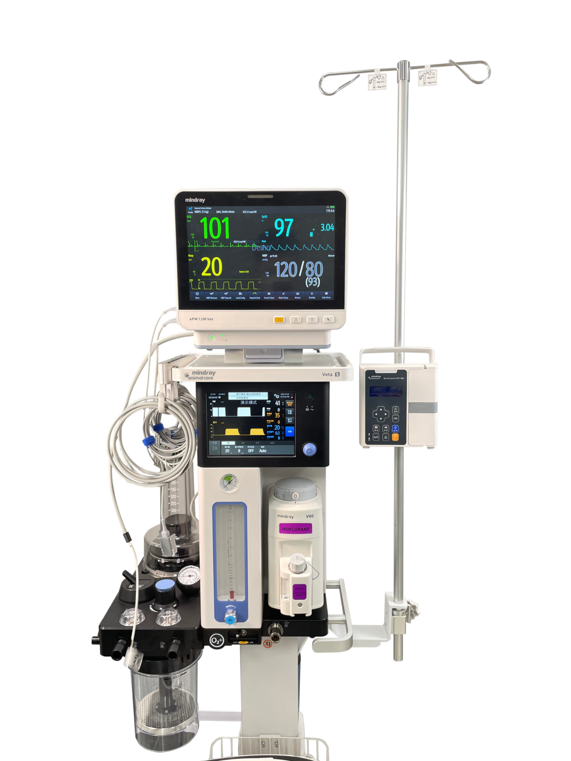
Our modern veterinary services include advanced diagnostic imaging, which X-ray and ultrasound in dogs, to ensure a comprehensive and accurate examination of your four-legged friends. These examinations often complement each other and give our vets the opportunity to make precise diagnoses and offer optimal treatment with state-of-the-art equipment of the highest quality.
Table of contents
X-ray in a dog
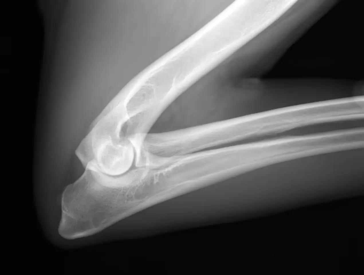
X-ray in a dog is a valuable tool for diagnosing and treating various health problems in our four-legged friends. X-rays provide a detailed picture of internal structures such as bones, organs and joints, thus providing insight that would otherwise be unavailable. At A-Vet, we use X-rays to examine both the skeleton and internal organs in the dog's chest and abdominal cavity. We offer advanced digital direct X-rays, which provide instant images and help detect conditions such as broken bones and the presence of foreign bodies in the abdomen. Our veterinarians are dedicated to using this technology to ensure thorough examinations.
How does the X-ray work?
X-ray is an imaging technology that uses X-rays, a type of electromagnetic radiation, to pass through the part of the dog's body to be examined. Bones and dense structures absorb more X-rays and therefore appear whiter in the images, while softer tissues such as organs and muscles let more rays through and appear darker. This creates a high-contrast image that gives us detailed information about the dog's internal anatomy.
Preparations for X-ray examination of dogs
Before an X-ray examination begins, the dog must undergo a thorough preparation to ensure accurate and reliable results. The vet will often ask the owners to fast the dog for a certain period before the examination, especially if the stomach or intestinal area is to be examined. This reduces the amount of air and food in the stomach, which improves the quality of the X-rays.
Depending on the dog's temperament, size and the specific area to be examined, it may be necessary to administer anesthesia or sedatives. This ensures that the dog remains calm and in a stable position throughout the procedure. A relaxed state is essential to avoid blurry images.
How is the X-ray examination carried out?
When the dog is ready, it will be placed on the x-ray table and the vet will adjust its position to get the most informative images. Often it requires several angles and images to get a complete picture of the desired area. In addition, we ensure minimal exposure to limit radiation risk.
After the pictures are taken, the vet will carefully analyze them to identify any abnormal findings. The results provide valuable information that can be used to make accurate diagnoses and develop an appropriate treatment plan.
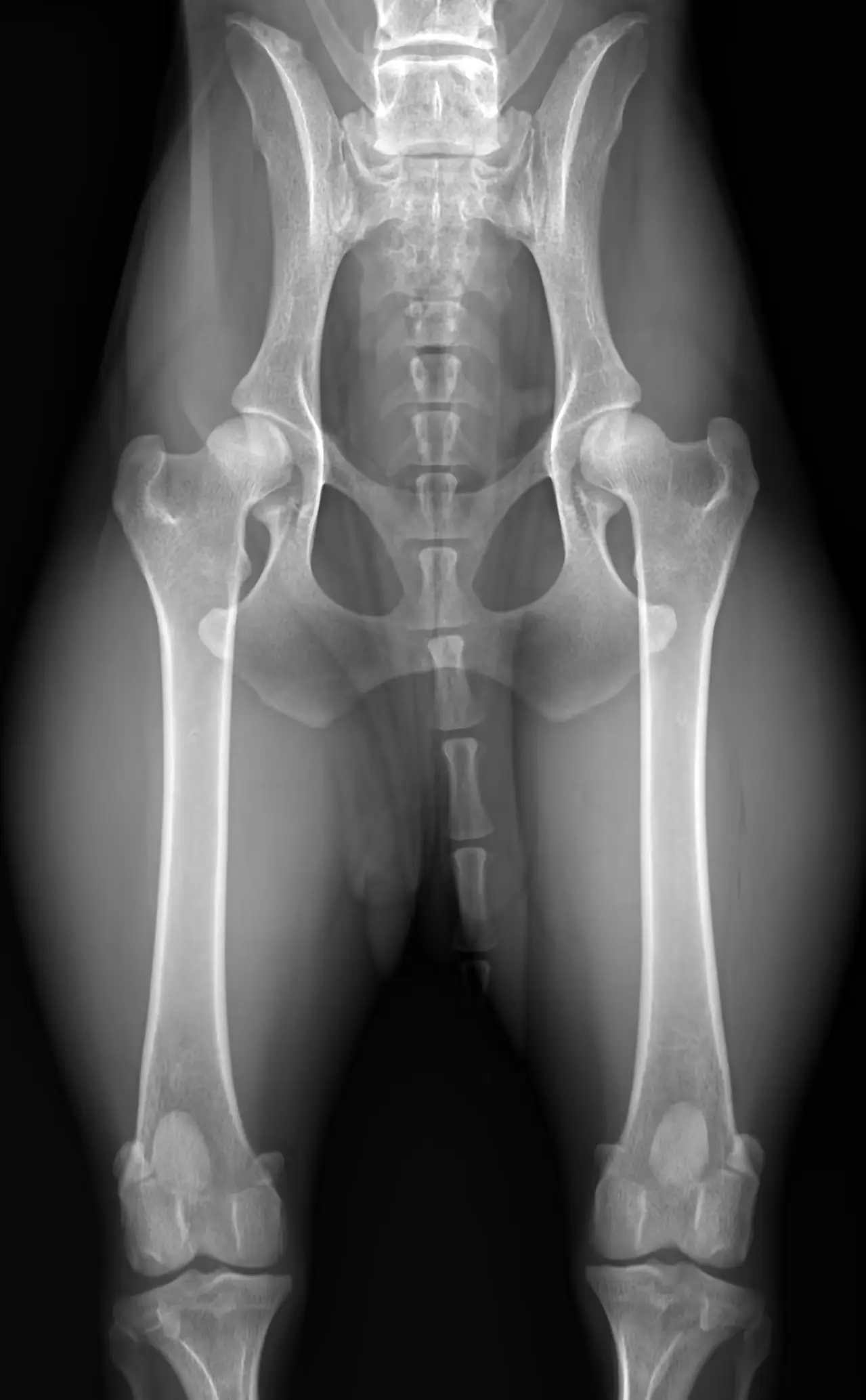
HD X-ray of dog
At A-Vet, we offer HD X-rays of dogs. Hip dysplasia (HD) is a hereditary developmental disorder that occurs in dogs of many breeds, where the hip socket and femoral head are malformed and do not fit together. This maldevelopment is rarely painful in itself, but it can lead to potentially painful sequelae.
Although all dogs can develop HD, it is most common in medium and large dog breeds. In order to gain an overview of and reduce the incidence of the disease, a large international effort is being made to screen for HD among dogs. This means that an X-ray is taken of a large number of dogs when they are over one year old. The images are then sent to a reading panel that reads the images and assesses the degree of HD based on international criteria. HD x-rays must be taken at a vet who is approved and has an agreement for this through NKK. This approval implies that the veterinarian has undergone a separate HD and AD course under the auspices of Norwegian Kennel Club og The Veterinary Association. All our veterinarians at A-Vet Small Animal Clinic are approved and have an agreement with the NKK.
Ultrasound in dogs
Ultrasound in dogs is an effective method for diagnosing and monitoring the health of our four-legged friends. This advanced method allows our veterinarians to examine internal organs without the need for complicated procedures, and it proves to be a gentle and reliable method of detecting various health problems in dogs. At A-Vet, we often use ultrasound in dogs to examine pregnancy, but it can also be used to diagnose heart and abdominal organ disorders.
How does ultrasound work?
Ultrasound, or high frequency sound waves, is the foundation of this technology. When the device is placed on the dog's skin, it sends sound waves into the body. These waves are reflected by the tissues in the body and return as echoes to the device. A computer then interprets these echoes and creates detailed images of organs, tissues and structures in real time.
Preparations for an ultrasound examination of the dog
Before the ultrasound examination begins, it is common for the dog to fast for a couple of hours beforehand. This ensures that the stomach is empty, and gives the ultrasound technician a clear visual access to the internal organs. If an ultrasound examination of the urinary bladder is performed, it is best that it is full.
In some cases, it may be necessary to anesthetize the dog to ensure calmness and stability during the examination. This is especially common if your dog is restless or has difficulty staying in a certain position. The vet will assess the dog's condition and decide if anesthesia or sedatives are necessary.
How is the ultrasound examination carried out?
The ultrasound examination is carried out by one of our experienced veterinarians, who places the dog on an examination table when everything is ready, and carefully applies a special gel to the area to be examined. The gel helps to eliminate air pockets between the skin and the device, which ensures clear and accurate images. The device is then gently guided over the area while images are displayed on a screen in real time.
The organs usually examined include the liver, kidneys, spleen, bladder, and reproductive organs. The images generated by the ultrasound machine provide information about the organs' size, structure and any diseases. This enables the vet to detect potential problems such as cysts, tumors or inflammation.
One of the advantages of ultrasound examinations is that they are painless and gentle. The dog usually does not require sedation for this procedure, unless it is agitated or anxious. Some owners still choose to be present during the examination to reassure the animal.
Fetal diagnostics of dogs
Pregnancy examination is the most common reason for an ultrasound examination. Here, the pregnancy can be established with certainty no earlier than 23 days after mating, and in some cases the pregnancy can be determined earlier. It can still be difficult to count the number of fetuses with certainty only with the help of ultrasound, especially in large litters. To know the exact number of kittens, an X-ray examination can be carried out closer to birth.
Diagnoses using ultrasound
Ultrasound is also helpful for examinations of the abdomen, and the vet will be able to see abdominal organs, vessels and lymph nodes and map changes in, among other things, the liver, kidneys and uterus. During an ultrasound examination of the abdomen, the dog must as a rule be shaved on the abdomen. It is also common to use ultrasound for echocardiography, which is an examination of the heart.
Expert assessment with teleradiology
At A-vet, we have skilled employees with good specialist knowledge in diagnostic imaging. In addition, we have a collaboration with ultrasound specialists in Oslo-based Telebuddies who perform telemedicine. Their veterinarians help with difficult cases both in X-ray and ultrasound when there is a need for it.
X-ray and ultrasound for dog price
Feel free to contact us if you have any questions about X-ray and ultrasound examinations for your dog. Information on prices can be found here. We are here to ensure that your pets receive the best care and treatment they deserve.